Positron Emission Tomography with monolithic crystals

Internship
Type of Project: Experimental Project
Location: Donostia
Supervisors:
S. Roberto Soleti, Samuele Torelli
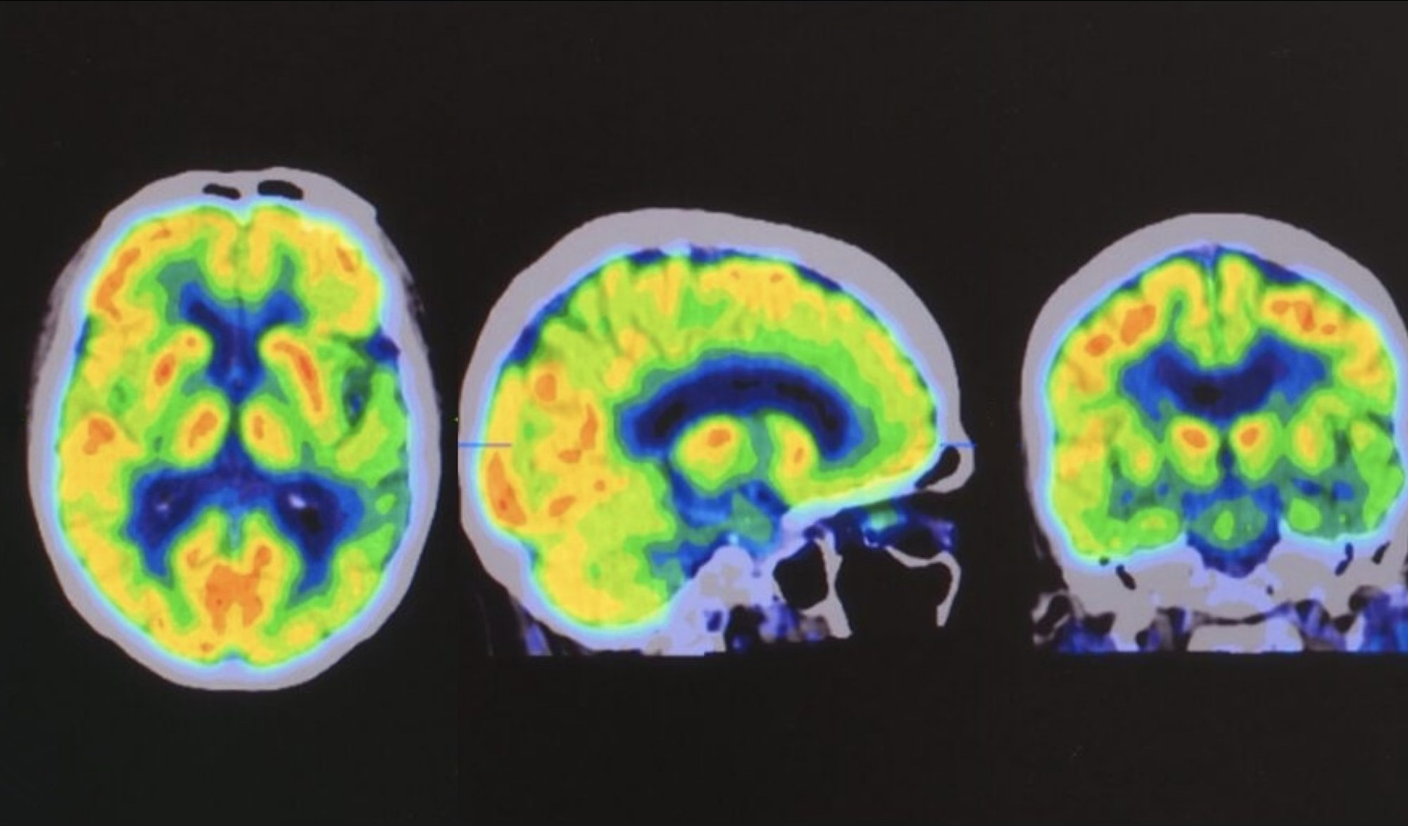
Positron-emission tomography (PET) is a powerful imaging technique that plays a pivotal role in medical diagnostics, utilizing positron-emitting radionuclides to track biologically active molecules. The annihilation of positrons and electrons produces gamma rays, which are detected to create detailed images of functional processes within the body.
Commercial PET scanners typically employ arrays of several thousand pixel-sized crystals. These crystals emit light when excited by ionizing radiation, such as gamma rays in a PET. In modern devices, the light emitted by the crystals is detected by silicon photomultipliers, which are efficient and robust solid-state photosensors. The dimensions of the pixel thus determine the spatial resolution of the device. In this project we will investigate the feasibility of using larger, monolithic crystals read out by an array of solid-state photosensors. This approach significantly simplifies the device’s design and assembly, reducing costs. While monolithic crystals typically face challenges in reconstructing the interaction point of the gamma radiation, recent advancements in machine learning algorithms for image processing could potentially enable the realization of a monolithic PET with performances analogous or better than a pixelated one.
This internship is an excellent opportunity to gain hands-on experience in a laboratory setting, working on a project that merges particle physics and medical imaging. The student will also have the chance to perform detailed microphysical simulations of the device and apply machine learning algorithms to reconstruct the interaction point of the gamma particle inside the crystal.
